See what the eye reveals
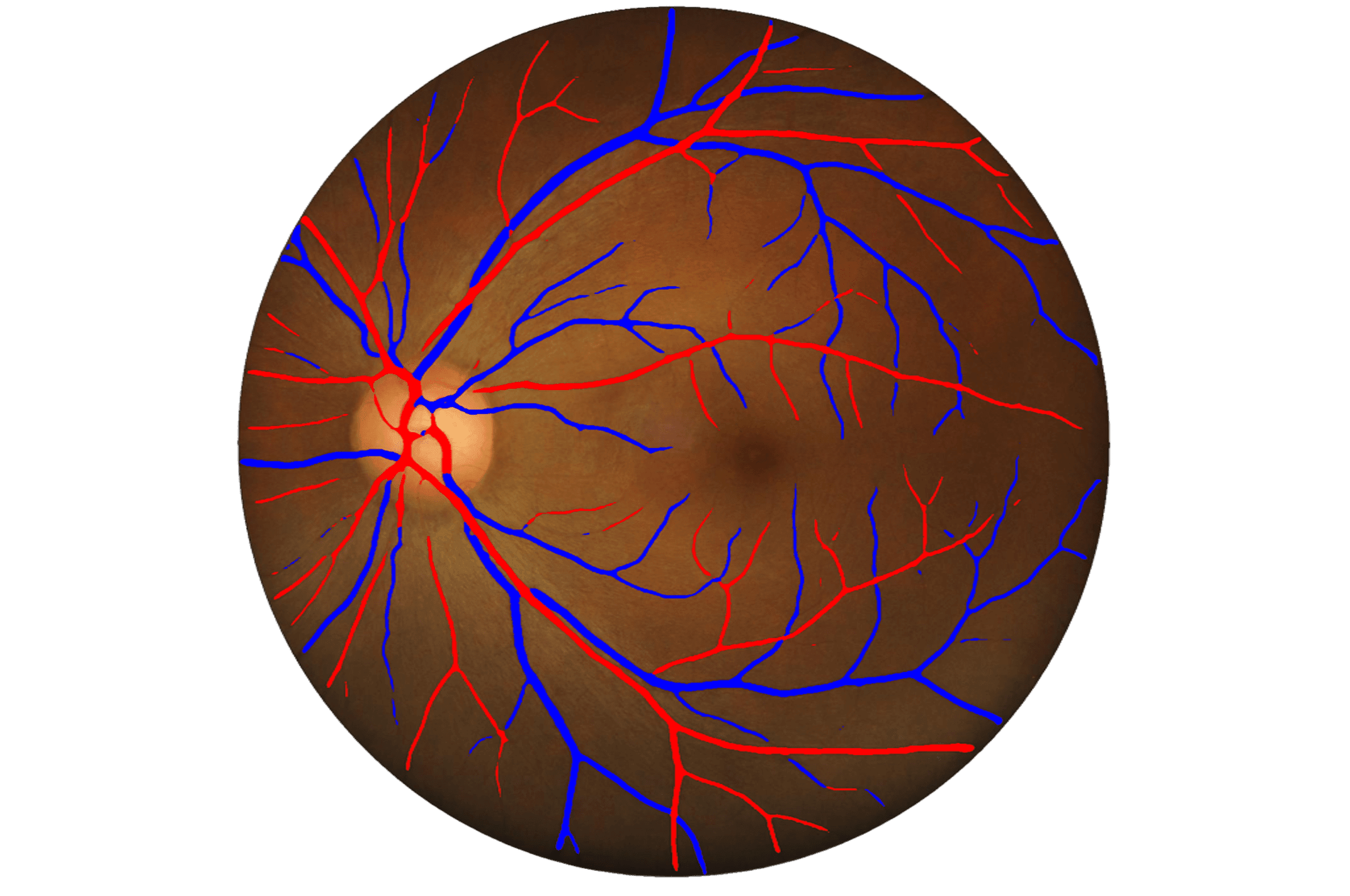
Benah AI transforms retinal images and videos into actionable diagnostic insights — helping ophthalmologists and clinicians detect and monitor retinal diseases with confidence and speed.
95%+
Detection accuracy
<30s
Per scan analysis
10+
Disease markers
*Based on Benah AI internal validation testing — see clinical research.
Built for clinical precision
Every feature in Benah AI is designed around the real constraints and requirements of clinical ophthalmology.
Multi-pathology Detection
Simultaneously screens for diabetic retinopathy, age-related macular degeneration, glaucoma, and more than ten additional retinal conditions in a single scan.
Image & Video Analysis
Processes both still fundus images and dynamic video recordings, capturing temporal changes and vascular patterns invisible in single frames.
Quantitative Grading
Produces severity scores, lesion counts, and longitudinal tracking measurements so clinicians can monitor disease progression over time.
Clinically Validated
Models trained and independently validated on diverse, multi-ethnic retinal datasets to ensure consistent performance across patient populations.
Seamless Integration
REST API and DICOM-compatible output make integration with existing PACS, EHR, and clinical management systems straightforward.
Results in Under 30 Seconds
Rapid inference means results arrive before the patient leaves the chair — enabling same-visit clinical decisions.
Patient & Practice Management
Built-in patient records, run history, and multi-user organisations — so your team can manage screening workflows without a separate practice-management tool.
Desktop & Mobile Capture Apps
Our desktop and Android apps connect to your Odocs fundus camera to capture, review, and submit images straight into the platform — already have one? No equipment to replace.
Making retinal expertise universally accessible
Hundreds of millions of people worldwide are at risk of vision-threatening retinal diseases, yet access to specialist ophthalmologists remains severely limited in many regions. Benah AI bridges this gap.
Our platform brings together advances in computer vision, clinical imaging, and medical AI to give every eye care professional — regardless of their location or resources — access to specialist-level retinal analysis.
Vision
A world where no retinal disease goes undetected
Mission
Precision AI tools that augment clinical expertise
For
Ophthalmologists, optometrists & clinicians globally
285M1
People living with vision impairment worldwide
80%1
Of blindness cases are preventable or treatable
10+2
Retinal conditions detectable with Benah AI
95%+2
Sensitivity in clinical validation studies
1. World Health Organization, Global Data on Visual Impairment. 2. Based on Benah AI internal validation testing.
From capture to clinical insight
Three seamless steps transform raw retinal data into clear, actionable reports for your diagnostic workflow.
Capture
Retinal image & video acquisition
Our desktop and mobile apps capture still images and video through your Odocs fundus camera. Already have one? No equipment to replace — and we can supply a camera or extend support to other devices if you need to.
Analyse
AI-powered computer vision
Our deep learning models detect and grade retinal pathologies across multiple disease categories — from diabetic retinopathy and macular degeneration to glaucomatous changes — in under 30 seconds.
Report
Structured outputs for experts
Clinicians receive annotated images, quantitative measurements, and a structured diagnostic report — all designed to support, not replace, the expert's final judgement.
Everything you need to start capturing
Benah AI's capture apps work with your Odocs fundus camera today. Don't have one yet, or use something else? We can help with that too.
Capture Apps
Our desktop and Android apps connect directly to your Odocs camera, letting your team capture, review, and submit images straight into the Benah AI platform.
Odocs Camera Sourcing
New to retinal imaging, or replacing an older camera? We can supply Odocs-compatible fundus cameras alongside your software setup, so your team has everything it needs from day one.
Custom Device Integration
Already using a different fundus camera or OCT device? Our team can extend Benah AI's compatibility to your existing equipment on request.
Support your diagnostic decisions with AI
Click a service to see an illustrative preview of Benah AI output.
How many patients screened this week have >2% papilledema probability?
3 patients this week have a papilledema probability above 2% — IDs #042, #118 and #156. #118 is the highest at 9.4% and may warrant urgent review.
Patient identifiers are stripped before any data reaches the AI model
For Clinical Research
Increase Research Site ROI with AI for OCT Scan Analysis
What you get
- Double the enrollment of eligible patients in clinical research
- Decrease screen failure rate
- Work with GA, Dry AMD, Wet AMD, DR, DME, RVO and other pathologies
- Monitor treatment using quantitative data: maps, graphs, %
- Analyze historical data of any volume and generate research-ready reports
- Generate clinical insights
- Analyze data from EMR/EHR systems
- Unlock vendor-neutrality: work with top 9 OCT devices
- Standardize endpoint data across sites
- Set higher standards across sites
Ready to see what Benah AI can do?
Start your free trial today and experience AI-powered retinal analysis integrated into your clinical workflow.
No credit card required · 30-day free trial · Cancel anytime
Let's talk about your clinic
Whether you're exploring Benah AI for a single practice or a large screening programme, our team is happy to help you find the right fit.